Calcaneal osteotomy represents one of the most transformative surgical interventions for patients suffering from complex foot deformities, particularly those with severe flatfoot conditions and hindfoot malalignment. This sophisticated surgical procedure involves the precise cutting and repositioning of the calcaneus (heel bone) to correct structural abnormalities that have developed over years or even decades. For individuals who have lived with progressive foot deformities, the decision to undergo calcaneal osteotomy often comes after conservative treatments have failed to provide adequate relief.
The journey toward surgical intervention frequently begins with years of adaptation and compromise. Many patients, like those experiencing severe flatfoot deformities, manage to maintain active lifestyles well into their fifth decade before the supporting structures of the foot begin to deteriorate significantly. When ligaments stretch beyond their capacity for self-repair and conservative measures no longer suffice, calcaneal osteotomy emerges as a viable solution for restoring both function and comfort to the affected foot.
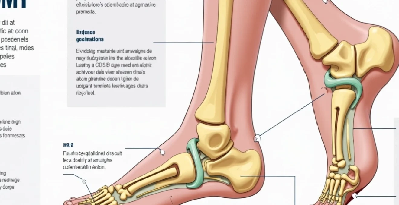
Understanding calcaneal osteotomy: dwyer and evans surgical techniques
The realm of calcaneal osteotomy encompasses several distinct surgical approaches, each designed to address specific deformity patterns and patient needs. These procedures have evolved considerably over the past several decades, with modern techniques offering improved outcomes and reduced complication rates compared to historical methods.
Lateral closing wedge dwyer osteotomy for hindfoot varus correction
The Dwyer osteotomy stands as one of the most established procedures for correcting hindfoot varus deformity. This technique involves removing a wedge-shaped piece of bone from the lateral aspect of the calcaneus, effectively allowing the heel to shift into a more anatomically correct position. The procedure addresses the fundamental issue of heel misalignment that contributes to pain, instability, and progressive deformity.
Surgeons typically recommend the Dwyer procedure when patients present with rigid hindfoot varus that cannot be corrected through conservative means. The lateral closing wedge technique offers precise control over the degree of correction achieved, allowing orthopaedic specialists to tailor the intervention to each patient’s specific anatomical requirements. Recovery from this procedure generally follows a predictable timeline, though individual variations in healing rates are common.
Medial opening wedge calcaneal lengthening evans procedure
The Evans calcaneal lengthening procedure addresses a different spectrum of foot deformities, particularly those involving forefoot abduction and midfoot collapse. This technique involves creating an osteotomy in the anterior aspect of the calcaneus and inserting a bone graft or synthetic wedge to lengthen the lateral column of the foot. The procedure effectively restores the foot’s natural arch configuration and improves overall biomechanical function.
Patients undergoing the Evans procedure often experience significant improvements in their ability to participate in weight-bearing activities. The technique has proven particularly effective for individuals with adult-acquired flatfoot deformity who have maintained reasonable subtalar joint mobility. The calcaneal lengthening approach provides excellent long-term outcomes when combined with appropriate soft tissue procedures and comprehensive rehabilitation protocols.
Z-cut calcaneal osteotomy for complex deformity reconstruction
Complex foot deformities sometimes require more sophisticated surgical approaches, and the Z-cut calcaneal osteotomy represents one such advanced technique. This procedure involves creating a Z-shaped cut through the calcaneus, allowing for multi-planar correction of deformity patterns. The technique proves particularly valuable when addressing combined deformities that involve both sagittal and coronal plane abnormalities.
The Z-cut approach offers surgeons greater flexibility in achieving comprehensive deformity correction while maintaining adequate bone contact for healing. Patients who undergo this procedure typically present with more severe deformity patterns and may require extended recovery periods compared to those undergoing simpler osteotomy techniques. The multi-planar correction capability of the Z-cut procedure makes it an invaluable tool in the orthopaedic surgeon’s armamentarium.
Plantarflexory osteotomy techniques for calcaneal pitch adjustment
Calcaneal pitch abnormalities can significantly impact foot function and patient comfort, necessitating specific surgical interventions designed to restore normal heel positioning. Plantarflexory osteotomy techniques focus on adjusting the angle at which the calcaneus meets the ground, effectively modifying the foot’s weight-bearing characteristics. These procedures prove particularly beneficial for patients with cavus foot deformities or those experiencing excessive heel elevation.
The success of plantarflexory osteotomy procedures depends largely on accurate pre-operative planning and precise surgical execution. Surgeons must carefully calculate the degree of correction required to achieve optimal post-operative alignment while avoiding overcorrection that could lead to secondary complications. Recovery from these procedures typically involves graduated weight-bearing protocols and specialised rehabilitation programmes.
Pre-operative assessment and surgical planning protocols
Comprehensive pre-operative evaluation forms the foundation of successful calcaneal osteotomy outcomes. The assessment process extends far beyond simple clinical examination, incorporating advanced imaging studies and biomechanical analyses to develop individualised surgical plans. Modern pre-operative protocols emphasise the importance of understanding each patient’s unique deformity pattern and functional requirements.
Weight-bearing radiographic analysis and meary’s angle measurement
Weight-bearing radiographic studies provide crucial information about foot alignment and deformity magnitude under physiological loading conditions. The measurement of Meary’s angle, which quantifies the relationship between the first metatarsal and the talus, serves as a fundamental parameter in assessing flatfoot severity. Normal Meary’s angle values typically range from 0 to 5 degrees, with greater angles indicating progressive midfoot collapse.
Advanced radiographic techniques now incorporate digital measurement tools that enhance accuracy and reproducibility in deformity assessment. These measurements directly influence surgical decision-making, helping surgeons determine the appropriate osteotomy technique and the degree of correction required. Quantitative radiographic analysis has significantly improved the predictability of surgical outcomes in calcaneal osteotomy procedures.
CT scan evaluation for subtalar joint arthritis assessment
Computed tomography scanning plays an essential role in evaluating the condition of the subtalar joint and surrounding bony structures. The presence of arthritis or joint space narrowing significantly influences surgical planning and may necessitate additional procedures such as subtalar joint fusion. CT imaging provides three-dimensional visualisation of bone architecture that cannot be appreciated through conventional radiography alone.
The identification of osteochondral defects, bone cysts, or other pathological changes through CT scanning helps surgeons anticipate potential complications and modify surgical approaches accordingly. Patients with significant subtalar arthritis may require combined procedures that address both the calcaneal deformity and joint pathology simultaneously. The three-dimensional imaging capability of CT scans has revolutionised pre-operative planning for complex foot reconstruction procedures.
Hindfoot alignment view and calcaneal pitch angle determination
The hindfoot alignment view represents a specialised radiographic projection that provides critical information about heel positioning in the coronal plane. This imaging study helps quantify the degree of hindfoot varus or valgus deformity and guides surgical correction strategies. Normal hindfoot alignment typically demonstrates a straight relationship between the tibia and calcaneus, with deviations indicating the need for corrective intervention.
Calcaneal pitch angle measurement complements the hindfoot alignment assessment by providing information about sagittal plane deformity. The pitch angle, measured between the calcaneus and the horizontal plane, normally ranges from 20 to 30 degrees. Significant deviations from this range indicate the need for sagittal plane correction during calcaneal osteotomy procedures. These measurements collectively inform surgical planning and help predict post-operative outcomes.
Silfverskiöld test and gastrocnemius contracture evaluation
The Silfverskiöld test provides valuable information about gastrocnemius and Achilles tendon contracture, conditions that frequently accompany calcaneal deformities. This clinical examination technique involves assessing ankle dorsiflexion range of motion with the knee in both extended and flexed positions. Significant differences between these measurements indicate isolated gastrocnemius contracture that may require concurrent surgical release.
Gastrocnemius contracture can significantly impact the success of calcaneal osteotomy procedures, as persistent equinus deformity may lead to recurrent symptoms or failure of deformity correction. The identification of contracture through the Silfverskiöld test allows surgeons to plan appropriate soft tissue releases that complement the bony correction. Combined approaches addressing both skeletal and soft tissue abnormalities generally produce superior long-term outcomes compared to isolated procedures.
Post-operative recovery timeline and rehabilitation milestones
The post-operative recovery process following calcaneal osteotomy represents a carefully orchestrated progression through distinct phases, each with specific goals and limitations. Understanding this timeline proves essential for patients preparing for surgery, as realistic expectations significantly influence satisfaction with surgical outcomes. The recovery process typically extends over 12 months, with major milestones occurring at predictable intervals throughout this period.
Non-weight-bearing phase: weeks 1-6 immobilisation protocol
The initial post-operative period demands strict adherence to non-weight-bearing protocols to ensure proper healing of the osteotomy site. During the first six weeks, patients typically remain in a cast or protective boot while maintaining complete avoidance of weight-bearing on the affected extremity. This phase often proves most challenging for active individuals accustomed to maintaining busy lifestyles and regular exercise routines.
Pain management during this period typically involves a combination of prescription medications, elevation, and ice therapy. Many patients report pain levels ranging from 1-2 at rest to 3-5 during repositioning or therapy activities. The immobilisation requirements necessitate significant lifestyle modifications, including reliance on assistive devices such as crutches or knee scooters for mobility. Patients often find that tasks previously taken for granted, such as carrying items or navigating stairs, require careful planning and adaptation.
The non-weight-bearing phase represents the most critical period for ensuring successful bone healing and optimal long-term outcomes following calcaneal osteotomy procedures.
Partial Weight-Bearing transition: weeks 7-12 progression guidelines
The transition to partial weight-bearing typically begins around the seventh post-operative week, contingent upon radiographic evidence of healing progression. This phase involves gradual introduction of weight-bearing forces while continuing to use protective footwear or orthotic devices. Patients often experience significant psychological relief during this period as mobility restrictions begin to ease and independence gradually returns.
Physical therapy typically commences during this phase, focusing on range of motion exercises, gait training, and gradual strengthening activities. The progressive loading protocol must be carefully monitored to avoid complications while encouraging appropriate tissue adaptation. Many patients report that while walking becomes possible, activities requiring prolonged standing or walking distances remain challenging during this period. Swelling and stiffness commonly persist, particularly after periods of increased activity.
Full Weight-Bearing achievement: month 3-4 activity resumption
The achievement of full weight-bearing status typically occurs between the third and fourth post-operative months, representing a significant milestone in the recovery process. At this stage, patients can generally discontinue assistive devices and begin wearing regular footwear, though supportive shoes remain recommended. The ability to return to work and resume many daily activities often coincides with this phase of recovery.
However, full weight-bearing does not immediately translate to unrestricted activity participation. Patients must continue avoiding high-impact activities and sports participation while tissues continue to adapt and strengthen. The gradual activity progression during this period helps prevent overuse injuries and ensures continued healing of both bone and soft tissue structures. Many individuals find that while they can walk normally, activities such as prolonged standing or walking on uneven surfaces may still cause discomfort or fatigue.
Return to sport and High-Impact activities: 6-month recovery benchmark
The return to sporting activities and high-impact exercises typically begins around the six-month post-operative mark, though individual variations in healing rates may influence this timeline. Patients eager to resume activities such as running, tennis, or other recreational sports must undergo careful evaluation to ensure adequate healing and functional recovery before clearance is granted.
The success of return to sport activities often depends on comprehensive rehabilitation programmes that address strength, flexibility, and proprioceptive deficits that may have developed during the recovery period. Many patients find that while they can successfully return to their desired activities, they may need to modify training routines or incorporate additional supportive measures such as custom orthotic devices or specialised footwear. The goal of returning to activities such as 5-kilometre runs or competitive tennis without subsequent pain or swelling represents the ultimate measure of surgical success for many individuals.
Common complications and management strategies in calcaneal osteotomy
Despite careful surgical technique and appropriate post-operative management, calcaneal osteotomy procedures carry inherent risks that patients must understand before proceeding with surgery. Complication rates vary depending on the specific technique employed, patient factors, and adherence to post-operative protocols. Modern surgical techniques have significantly reduced complication rates compared to historical approaches, though certain risks persist and require vigilant monitoring throughout the recovery process.
Delayed union or nonunion represents one of the most significant potential complications following calcaneal osteotomy. This condition occurs when the bone fails to heal adequately within the expected timeframe, potentially requiring additional surgical intervention. Risk factors for healing complications include smoking, diabetes, poor nutritional status, and non-compliance with weight-bearing restrictions. Bone healing complications can significantly extend recovery timelines and may necessitate bone grafting procedures or revision surgery to achieve satisfactory outcomes.
Infection remains a concern in any surgical procedure, though the risk is relatively low in calcaneal osteotomy when appropriate prophylactic measures are employed. Superficial infections typically respond well to antibiotic therapy, while deep infections may require surgical debridement and extended antibiotic courses. Patients can minimise infection risk through proper wound care, adherence to activity restrictions, and prompt reporting of concerning signs such as increased pain, swelling, or drainage from the surgical site.
Overcorrection or undercorrection of the intended deformity represents another potential complication that can significantly impact functional outcomes. Precise pre-operative planning and careful surgical technique help minimise this risk, though anatomical variations and healing responses can sometimes lead to suboptimal alignment. Residual deformity or overcorrection may require additional procedures to achieve satisfactory functional and cosmetic results, highlighting the importance of realistic expectations and thorough pre-operative counselling.
Understanding potential complications and their management strategies enables patients to make informed decisions about surgical intervention while maintaining realistic expectations about recovery outcomes.
Long-term outcomes and functional assessment measures
The evaluation of long-term outcomes following calcaneal osteotomy requires comprehensive assessment of both objective functional measures and subjective patient satisfaction indicators. Research studies consistently demonstrate that the majority of patients experience significant improvement in pain levels, functional capacity, and quality of life measures following successful calcaneal osteotomy procedures. However, individual outcomes can vary considerably based on pre-operative deformity severity, surgical technique, and patient compliance with rehabilitation protocols.
Functional outcome measures typically include assessment of walking distance, stair climbing ability, return to recreational activities, and overall satisfaction with surgical results. Studies indicate that approximately 80-90% of patients report significant improvement in pain levels and functional capacity at two-year follow-up appointments. The ability to return to activities such as recreational running, tennis, or hiking represents a key success indicator for many patients who underwent surgery specifically to resume these pursuits.
Patient-reported outcome measures (PROMs) provide valuable insights into the subjective experience of recovery and long-term satisfaction with surgical results. These standardised questionnaires assess domains such as pain, function, and quality of life both before and after surgical intervention. Long-term follow-up studies demonstrate that satisfaction rates generally remain high even at five and ten-year intervals, suggesting that successful calcaneal osteotomy provides durable improvement in patient outcomes.
The development of secondary arthritis in adjacent joints represents a potential long-term concern following calcaneal osteotomy procedures. While the corrected alignment generally reduces abnormal stress on surrounding joints, some patients may develop degenerative changes over time, particularly in the subtalar or midfoot joints. Regular long-term follow-up allows for early detection and management of these secondary conditions, helping to preserve overall foot function and patient satisfaction throughout the years following surgery.
Return to high-level athletic activities remains possible for
many patients who maintain realistic expectations about the recovery process and commit to appropriate rehabilitation programmes. However, the timeline for returning to competitive or high-intensity activities may extend beyond the typical six-month benchmark, particularly for sports requiring rapid directional changes or sustained high-impact loading patterns.
Patient experience: personal recovery journey and adaptive strategies
The personal journey through calcaneal osteotomy recovery often proves more challenging than patients initially anticipate, despite thorough pre-operative counselling and preparation. Real-world experiences frequently differ from clinical descriptions, with individual healing patterns, pain tolerance levels, and lifestyle factors all contributing to unique recovery trajectories. Understanding these personal experiences provides valuable insights for prospective patients considering similar surgical interventions.
Many patients describe the initial weeks following surgery as a period of significant lifestyle adjustment, requiring careful planning and adaptation strategies for even basic daily activities. The transition from an active lifestyle to mobility dependence often creates psychological challenges that extend beyond the physical aspects of recovery. Emotional resilience becomes as important as physical healing during this period, with many individuals benefiting from maintaining realistic expectations while focusing on incremental progress markers.
The experience of navigating daily challenges while wearing protective boots or casts reveals the importance of environmental considerations that may not be immediately apparent during pre-operative planning. Simple tasks such as descending stairs can become significant obstacles, particularly when architectural features like interrupted handrails or non-functioning lifts compound the difficulty. Patients often develop creative problem-solving strategies, such as backwards stair descent or alternative route planning, to maintain independence during the recovery period.
The journey through calcaneal osteotomy recovery teaches patients valuable lessons about patience, adaptation, and the gradual nature of healing that extends far beyond the surgical site itself.
Long-term success stories from patients who have achieved their pre-operative goals provide encouragement for those currently navigating the recovery process. Individuals who successfully return to activities such as 5-kilometre running, competitive tennis, or recreational hiking often report that the extended recovery period ultimately proved worthwhile despite the initial challenges. These success stories typically emphasise the importance of adhering to rehabilitation protocols, maintaining realistic timeline expectations, and celebrating incremental improvements throughout the healing process.
The development of adaptive strategies during recovery often leads to improved overall foot health awareness and injury prevention practices that benefit patients long after complete healing has occurred. Many individuals report developing better understanding of proper footwear selection, the importance of gradual activity progression, and recognition of early warning signs that might indicate the need for activity modification. Post-surgical lifestyle modifications frequently include continued use of supportive footwear, regular stretching routines, and ongoing attention to foot biomechanics that contribute to sustained positive outcomes.
Patient testimonials consistently highlight the importance of having realistic expectations about the recovery timeline, particularly regarding the return to high-impact activities and sports participation. While some individuals achieve remarkable functional recovery within the expected timeframes, others require extended periods before reaching their optimal functional capacity. The variability in individual healing responses underscores the importance of patient education and the need for ongoing communication between patients and their surgical teams throughout the recovery process.
The psychological aspects of recovery often prove as significant as the physical components, with many patients experiencing periods of frustration, anxiety, or doubt about their decision to proceed with surgery. Support from family members, healthcare providers, and others who have undergone similar procedures can prove invaluable during challenging phases of recovery. Online communities and patient forums frequently provide practical advice and emotional support that complement formal medical care throughout the healing journey.
Successful long-term outcomes typically result from a combination of appropriate surgical technique, patient compliance with post-operative instructions, and realistic expectations about the recovery process. Patients who achieve their goals of pain-free activity participation often describe the surgery as life-changing, enabling them to return to beloved activities that had become impossible due to progressive foot deformity. The ability to walk, run, or participate in sports without subsequent pain or swelling represents the ultimate measure of surgical success for most individuals who undergo calcaneal osteotomy procedures.