The confusion between vitamin B12 and iron represents one of the most common misconceptions in nutritional health. Both nutrients play crucial roles in preventing anaemia and maintaining optimal blood health, yet they operate through entirely different biochemical mechanisms. Understanding these distinctions becomes particularly important when addressing deficiency-related conditions, as misidentifying the underlying cause can lead to inappropriate treatment approaches. This misconception often arises because both deficiencies can manifest similar symptoms, including fatigue, weakness, and various forms of anaemia, creating diagnostic challenges for healthcare practitioners and patients alike.
Fundamental biochemical differences between vitamin B12 and iron
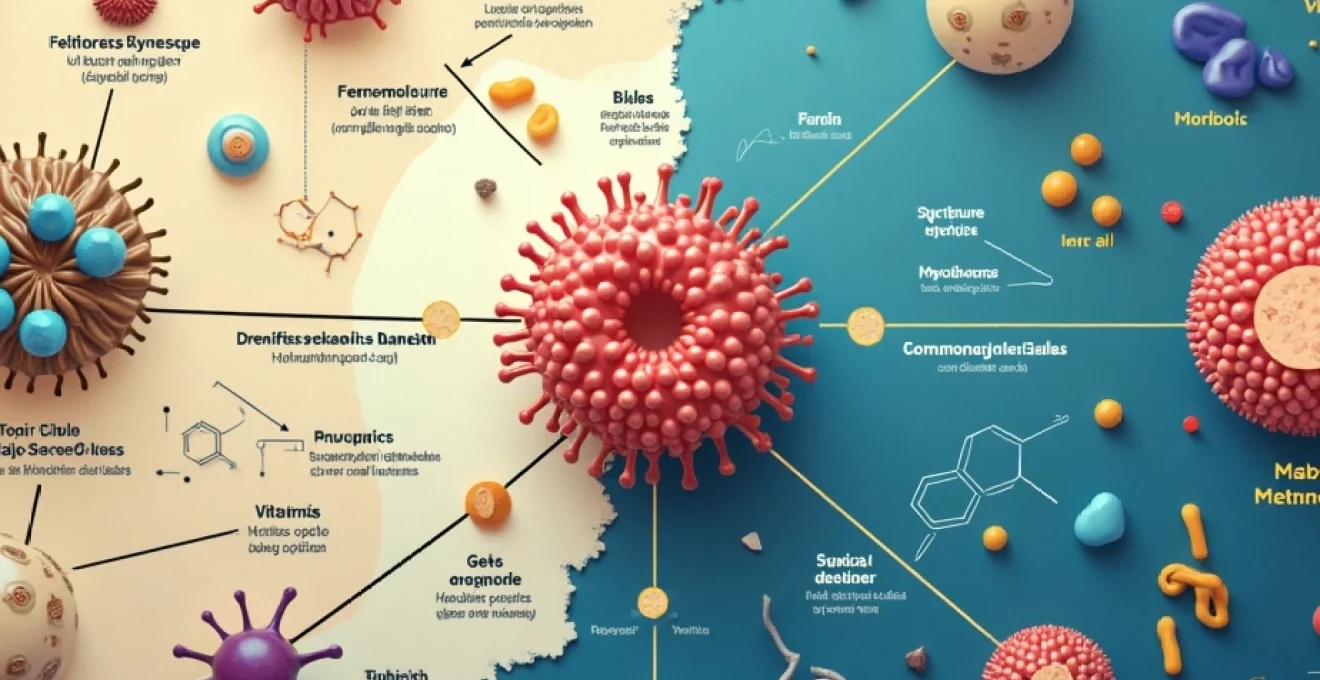
The structural and chemical differences between vitamin B12 and iron highlight their distinct biological roles. These two essential nutrients operate through completely separate metabolic pathways, despite both contributing to blood health and cellular function.
Cobalamin structure and Cobalt-Centred corrin ring chemistry
Vitamin B12, scientifically known as cobalamin, represents one of the most complex vitamins in human biochemistry. Its molecular structure features a distinctive cobalt atom positioned at the centre of a corrin ring system, surrounded by four nitrogen atoms in a square planar arrangement. This unique architecture allows B12 to participate in essential methylation reactions and DNA synthesis processes. The corrin ring differs significantly from the porphyrin rings found in other biological molecules, making cobalamin structurally unique among vitamins. Various forms of B12 exist, including cyanocobalamin, methylcobalamin, and adenosylcobalamin, each serving specific physiological functions.
Iron’s role as a transition metal in haemoglobin and myoglobin
Iron functions as a transition metal with variable oxidation states, primarily existing as ferrous (Fe²⁺) and ferric (Fe³⁺) forms in biological systems. Within haemoglobin molecules, iron atoms coordinate with nitrogen atoms in porphyrin rings, creating the haem group responsible for oxygen binding and transport. This metal’s ability to reversibly bind oxygen makes it indispensable for cellular respiration. Iron also plays critical roles in myoglobin for oxygen storage in muscles, and in various enzymes including cytochromes involved in electron transport chains. The metal’s electron configuration allows for efficient electron transfer reactions, supporting numerous metabolic processes.
Molecular weight disparities: cyanocobalamin vs ferrous sulphate
The molecular weight differences between these nutrients reflect their structural complexity. Cyanocobalamin, the most stable form of vitamin B12, has a molecular weight of approximately 1,355 daltons, making it one of the largest vitamins. In contrast, elemental iron has an atomic weight of just 55.8 daltons, while common iron supplements like ferrous sulphate weigh 151.9 daltons. This substantial size difference influences absorption mechanisms, with B12 requiring specific transport proteins due to its large molecular structure. The complexity of B12’s molecular architecture necessitates intricate absorption processes , unlike iron’s relatively straightforward uptake mechanisms.
Water-soluble vitamin classification vs essential mineral element
Vitamin B12 belongs to the water-soluble B-complex vitamin family, though it exhibits unique characteristics among this group. Unlike other water-soluble vitamins that require frequent replenishment, B12 can be stored in the liver for several years. Iron, conversely, represents an essential mineral element that the body carefully regulates through absorption control mechanisms. The body maintains iron homeostasis through hepcidin regulation, preventing both deficiency and toxicity. These fundamental classification differences affect dietary requirements, storage capabilities, and excretion patterns between the two nutrients.
Distinct physiological functions and metabolic pathways
Understanding the separate metabolic roles of vitamin B12 and iron reveals why both nutrients remain essential despite serving different physiological functions. Their distinct pathways demonstrate the complexity of human metabolism and the importance of adequate intake of both nutrients.
Vitamin b12’s role in Methylmalonyl-CoA mutase activity
Vitamin B12 serves as a cofactor for methylmalonyl-CoA mutase, an enzyme critical for odd-chain fatty acid metabolism and amino acid catabolism. This enzyme converts methylmalonyl-CoA to succinyl-CoA, allowing these molecules to enter the citric acid cycle for energy production. B12 deficiency leads to methylmalonic acid accumulation, which serves as a diagnostic marker for cobalamin insufficiency. The vitamin also functions as a cofactor for methionine synthase, facilitating homocysteine conversion to methionine and supporting DNA methylation processes. These methylation reactions prove essential for proper gene expression and cellular function , highlighting B12’s role beyond simple blood formation.
Iron-dependent oxygen transport via haemoglobin synthesis
Iron’s primary physiological function centres on oxygen transport through haemoglobin synthesis within erythrocytes. Each haemoglobin molecule contains four iron atoms capable of binding oxygen molecules reversibly, enabling efficient oxygen delivery from lungs to tissues. Iron also supports myoglobin function in muscle tissues, providing oxygen storage for periods of high metabolic demand. Beyond oxygen transport, iron participates in electron transport chain complexes within mitochondria, supporting cellular energy production. The metal’s involvement in various enzymatic reactions, including those required for collagen synthesis and neurotransmitter production, demonstrates its broad physiological significance.
Intrinsic Factor-Mediated B12 absorption in terminal ileum
Vitamin B12 absorption requires intrinsic factor, a glycoprotein produced by gastric parietal cells, creating one of the most complex vitamin absorption mechanisms in human physiology. The B12-intrinsic factor complex travels through the digestive system to the terminal ileum, where specific receptors facilitate uptake. This process can take several hours and involves multiple transport proteins, including transcobalamin II for cellular delivery. Pernicious anaemia occurs when autoimmune destruction of parietal cells eliminates intrinsic factor production, preventing B12 absorption despite adequate dietary intake. This sophisticated absorption mechanism explains why B12 deficiency can develop even with sufficient dietary consumption .
Transferrin-bound iron uptake and ferritin storage mechanisms
Iron absorption occurs primarily in the duodenum through divalent metal transporter 1 (DMT1) and involves reduction from ferric to ferrous forms by duodenal cytochrome b reductase. Once absorbed, iron binds to transferrin for transport throughout the body, with transferrin saturation serving as an important diagnostic marker. Cellular iron uptake occurs through transferrin receptors, particularly abundant in rapidly dividing cells requiring iron for DNA synthesis. Excess iron stores as ferritin within cells, particularly in the liver, spleen, and bone marrow. This storage mechanism prevents iron toxicity while maintaining reserves for periods of increased demand or decreased intake.
Clinical deficiency syndromes and diagnostic markers
The clinical presentations of vitamin B12 and iron deficiencies differ significantly, providing healthcare professionals with distinct diagnostic patterns. Understanding these differences enables appropriate testing strategies and targeted treatment approaches for each condition.
Pernicious anaemia and megaloblastic cell morphology
Vitamin B12 deficiency typically manifests as megaloblastic anaemia, characterised by the production of abnormally large, immature red blood cells called megaloblasts. These cells result from impaired DNA synthesis due to inadequate B12 availability for methylation reactions. Pernicious anaemia, the most common cause of B12 deficiency in developed countries, develops when autoimmune destruction of gastric parietal cells eliminates intrinsic factor production. Blood smears reveal hypersegmented neutrophils and oval macrocytes, distinctive morphological features absent in iron deficiency. The condition often progresses slowly, allowing patients to adapt to gradually declining haemoglobin levels before symptoms become apparent.
Megaloblastic anaemia represents a fundamental disruption in DNA synthesis, creating distinctly abnormal cellular morphology that differentiates B12 deficiency from other anaemic conditions.
Iron-deficiency anaemia and microcytic hypochromic erythrocytes
Iron deficiency produces microcytic hypochromic anaemia, featuring small, pale red blood cells with reduced haemoglobin content. These morphological changes reflect the body’s inability to synthesise adequate haemoglobin due to insufficient iron availability. Early iron deficiency may present as iron depletion without anaemia, detectable only through ferritin measurements. As deficiency progresses, patients develop iron-deficient erythropoiesis, followed by overt anaemia. The condition typically develops more rapidly than B12 deficiency, particularly in cases involving blood loss. The microcytic pattern contrasts sharply with the macrocytic presentation of B12 deficiency , providing clear diagnostic differentiation.
Serum methylmalonic acid and homocysteine elevation in B12 deficiency
Elevated serum methylmalonic acid (MMA) serves as a highly sensitive marker for vitamin B12 deficiency, often increasing before serum B12 levels decline significantly. MMA accumulates when methylmalonyl-CoA mutase cannot function properly due to inadequate B12 availability. Homocysteine levels also rise in B12 deficiency due to impaired methionine synthase activity, though this marker lacks specificity as folate deficiency produces similar elevations. These metabolic markers prove particularly valuable when serum B12 levels appear borderline or when functional deficiency exists despite apparently normal B12 concentrations. MMA testing provides superior diagnostic accuracy compared to serum B12 measurements alone .
Ferritin, transferrin saturation, and total Iron-Binding capacity testing
Iron deficiency assessment relies on multiple laboratory parameters providing comprehensive evaluation of iron status. Serum ferritin represents the most sensitive single test for iron deficiency, reflecting total body iron stores. However, ferritin levels can be elevated by inflammation, infection, or liver disease, potentially masking concurrent iron deficiency. Transferrin saturation, calculated from serum iron and total iron-binding capacity measurements, indicates the percentage of transferrin molecules carrying iron. Low transferrin saturation suggests iron deficiency, while elevated total iron-binding capacity reflects increased transferrin production in response to iron depletion.
Pharmaceutical forms and bioavailability considerations
The pharmaceutical development of vitamin B12 and iron supplements reflects their distinct chemical properties and absorption mechanisms. Bioavailability considerations play crucial roles in determining appropriate dosing strategies and formulation choices for each nutrient. Understanding these pharmacological differences helps optimise therapeutic outcomes while minimising adverse effects.
Vitamin B12 supplements typically contain cyanocobalamin, the most stable synthetic form, or methylcobalamin, the naturally occurring active form. Cyanocobalamin requires conversion to active forms within the body, while methylcobalamin provides immediate biological activity. Sublingual and injectable formulations bypass the complex gastrointestinal absorption process, proving particularly valuable for patients with pernicious anaemia or gastrointestinal disorders. The large molecular size of B12 necessitates specific transport mechanisms, making bioavailability highly dependent on intrinsic factor availability and terminal ileum function. Injectable B12 formulations achieve 100% bioavailability , explaining their preference for treating severe deficiency states.
Iron supplements exist in multiple chemical forms, including ferrous sulphate, ferrous gluconate, and ferrous fumarate, each offering different elemental iron content and absorption characteristics. Ferrous forms demonstrate superior bioavailability compared to ferric formulations due to preferential uptake by duodenal transporters. Chelated iron forms, such as iron bisglycinate, may reduce gastrointestinal side effects while maintaining absorption efficiency. Enteric-coated formulations aim to reduce gastric irritation but may compromise absorption by delivering iron to intestinal segments with lower uptake capacity. The bioavailability of oral iron rarely exceeds 10-15% under optimal conditions, necessitating higher doses to meet therapeutic requirements.
Drug interactions and absorption interference mechanisms
Numerous medications and dietary factors influence the absorption and utilisation of both vitamin B12 and iron, though through entirely different mechanisms. Understanding these interactions proves essential for optimising therapeutic outcomes and preventing treatment failures in deficiency states.
Vitamin B12 absorption faces interference from proton pump inhibitors, H2 receptor antagonists, and metformin, which reduce gastric acid production or alter intrinsic factor function. Long-term use of these medications can gradually deplete B12 stores, particularly in older adults with already compromised absorption capacity. Nitrous oxide exposure can rapidly inactivate existing B12 stores, potentially precipitating acute deficiency in susceptible individuals. Alcohol consumption impairs B12 absorption through multiple mechanisms, including reduced intrinsic factor production and direct mucosal damage. The complexity of B12 absorption makes it vulnerable to numerous pharmaceutical and lifestyle interruptions .
Understanding medication-induced B12 and iron deficiencies enables proactive monitoring and supplementation strategies for at-risk patients.
Iron absorption encounters significant dietary and pharmaceutical interference, with calcium, tannins, and phytates forming insoluble complexes that prevent uptake. Concurrent administration of tetracycline antibiotics, quinolones, or bisphosphonates creates chelation complexes that reduce absorption of both iron and the medications. Proton pump inhibitors impair iron absorption by reducing gastric acid necessary for ferric-to-ferrous conversion. Vitamin C enhances iron absorption by maintaining iron in the ferrous state and forming absorbable complexes. The timing of iron supplementation relative to meals and other medications significantly influences bioavailability, often requiring careful scheduling to optimise therapeutic effectiveness.
Clinical applications in treating anaemia types
The clinical management of vitamin B12 and iron deficiency anaemias requires distinctly different therapeutic approaches, reflecting their separate pathophysiological mechanisms. Treatment success depends on accurate diagnosis, appropriate supplementation strategies, and addressing underlying causative factors. Healthcare providers must consider patient-specific factors, including age, comorbidities, and medication interactions, when developing treatment protocols.
Vitamin B12 deficiency treatment typically begins with high-dose injections to rapidly replenish depleted stores and bypass absorption limitations. Initial therapy often involves daily or alternate-day intramuscular injections for two weeks, followed by maintenance schedules based on the underlying cause. Patients with pernicious anaemia require lifelong treatment, while those with dietary deficiency may transition to oral supplementation once stores are repleted. Neurological complications necessitate more aggressive and prolonged treatment protocols, as nerve damage may be irreversible if not addressed promptly. The response to B12 treatment often occurs rapidly, with reticulocytosis beginning within days of initiation .
Iron deficiency anaemia treatment focuses on oral supplementation with elemental iron doses of 150-200mg daily, typically divided into multiple doses to enhance tolerance. Treatment duration usually extends 3-6 months beyond normalisation of haemoglobin levels to replenish iron stores adequately. Intravenous iron therapy becomes necessary for patients with severe anaemia, malabsorption, or intolerance to oral preparations. Monitoring includes regular assessment of haemoglobin, ferritin, and transferrin saturation to evaluate treatment response and guide therapy duration. Identifying and addressing underlying causes of blood loss remains crucial for preventing recurrence and ensuring sustained treatment success.
Combined deficiencies of both vitamin B12 and iron can occur simultaneously, particularly in patients with gastrointestinal disorders, dietary restrictions, or chronic diseases. These cases require careful diagnosis and treatment of both deficiencies, as correcting only one nutrient may mask the other deficiency or result in incomplete therapeutic response. Recent research suggests potential interactions between B12 and iron metabolism, indicating that adequate B12 status may optimise iron utilisation and vice versa. This emerging understanding emphasises the importance of comprehensive nutritional assessment and may influence future treatment guidelines for complex anaemic presentations.