Discovering a lump or bump on your shin bone can trigger immediate concern, particularly when you’re unsure whether it represents a benign condition or something more serious. The tibia, commonly known as the shin bone, is one of the most prominent bones in the human body and susceptible to various conditions that can manifest as palpable masses or protrusions. Understanding the difference between harmless developmental changes and potentially serious pathology becomes crucial for making informed decisions about seeking medical attention.
While many shin bone abnormalities stem from sports injuries, growth-related changes, or minor trauma, some presentations warrant immediate medical evaluation. The location, characteristics, and associated symptoms of tibial lumps provide valuable clues about their underlying nature. From common athletic injuries like shin splints to rare bone cancers such as adamantinoma, the spectrum of conditions affecting the tibia varies dramatically in both prevalence and clinical significance.
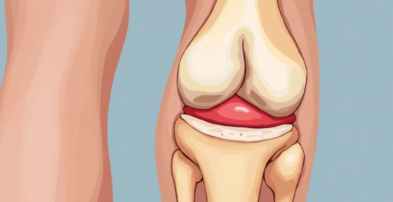
Anatomical structure and common locations of tibial lumps
The tibia serves as the primary weight-bearing bone of the lower leg, extending from the knee joint to the ankle. Its subcutaneous location makes it particularly vulnerable to direct trauma and creates conditions where abnormalities become readily apparent through visual inspection and palpation. The bone’s superficial position means that even minor changes in contour or the development of small masses can be easily detected by patients during routine activities.
Medial tibial stress syndrome and periosteal reactions
Medial tibial stress syndrome , commonly referred to as shin splints, represents one of the most frequent causes of shin discomfort and localised swelling. This condition develops when repeated stress causes inflammation of the periosteum, the thin membrane covering the bone surface. Athletes, particularly runners and dancers, experience this condition at higher rates due to repetitive impact loading on the tibial shaft.
The inflammatory response associated with medial tibial stress syndrome can create areas of tenderness and mild swelling along the posteromedial border of the tibia. While not typically forming discrete lumps, the periosteal thickening can create palpable irregularities that concern patients. The condition usually responds well to rest, ice application, and gradual return to activity with proper biomechanical assessment.
Osgood-schlatter disease in adolescent athletes
Osgood-Schlatter disease affects the anterior tibial tubercle, creating a prominent bump just below the knee cap in growing adolescents. This condition results from repeated traction on the developing tibial tuberosity by the powerful quadriceps muscle group during periods of rapid skeletal growth. Young athletes participating in sports requiring frequent jumping, running, or kicking movements face increased risk of developing this characteristic tibial prominence.
The condition typically manifests as a tender, often quite pronounced bump on the upper shin bone that becomes more noticeable during physical activity. Pain usually accompanies the visible swelling, particularly during knee extension activities. Most cases resolve spontaneously once skeletal maturity is achieved , though the bony prominence may persist throughout adult life as a harmless reminder of adolescent athletic participation.
Tibial tuberosity prominence and growth plate abnormalities
Variations in tibial tuberosity development can create concerning appearances for parents and young athletes. The tuberosity serves as the attachment point for the patellar tendon and experiences significant mechanical stress during the adolescent growth spurt. Some individuals develop more prominent tuberosities than others due to genetic factors, activity levels, and growth patterns.
Occasionally, growth plate irregularities can cause asymmetric development of the tibial tuberosity, leading to unusual contours or apparent masses. These developmental variations rarely require treatment but may necessitate imaging studies to differentiate them from pathological conditions. Understanding normal anatomical variation helps distinguish benign developmental changes from concerning pathology requiring intervention.
Anterior compartment syndrome manifestations
Chronic compartment syndrome affecting the anterior compartment of the leg can create swelling and firmness along the lateral aspect of the tibia. This condition develops when increased pressure within the fascial compartment compromises circulation and normal tissue function. Athletes engaged in repetitive activities may develop chronic symptoms that include muscle swelling and altered leg contour.
The condition typically presents with activity-related pain and swelling that subsides with rest. However, severe cases can develop persistent swelling that creates palpable changes in the normal leg contour. Compartment pressure measurements may be necessary to confirm the diagnosis and guide treatment decisions, which can range from conservative management to surgical fascial release.
Benign causes of shin bone protrusions
The majority of shin bone lumps encountered in clinical practice represent benign conditions that pose no threat to long-term health. These benign masses can arise from various tissues including bone, cartilage, and soft tissues adjacent to the tibia. Understanding the characteristics of common benign conditions helps patients and healthcare providers distinguish them from more concerning pathology requiring urgent intervention.
Exostosis formation and hereditary multiple exostoses
Osteochondromas, also known as exostoses, represent the most common benign bone tumours affecting the tibia. These cartilage-capped bony projections typically develop during childhood and adolescence, growing away from the joint and towards the middle of the bone shaft. While usually asymptomatic, larger exostoses can create cosmetic concerns or interfere with adjacent soft tissue structures.
Hereditary multiple exostoses is a genetic condition causing the development of numerous osteochondromas throughout the skeleton, including the tibia. Patients with this condition require regular monitoring due to a small risk of malignant transformation, particularly in larger lesions. The lifetime risk of malignant change remains relatively low , estimated at approximately 1-5% of affected individuals, but warrants ongoing surveillance throughout adult life.
Lipomas and soft tissue masses adjacent to tibia
Lipomas represent the most common soft tissue tumours encountered in clinical practice and can develop adjacent to the tibia, creating palpable masses that patients may interpret as bone-related abnormalities. These benign fatty tumours typically present as soft, mobile masses that remain painless unless they reach significant size or compress adjacent structures.
The superficial location of the tibia means that even small lipomas can become readily apparent to patients during self-examination. Most lipomas require no treatment unless they cause functional impairment or cosmetic concerns. Distinguishing lipomas from other soft tissue masses requires careful clinical evaluation and may necessitate imaging studies to confirm the diagnosis.
Ganglion cysts and synovial fluid accumulation
Ganglion cysts can occasionally develop near the tibia, particularly around the ankle region where multiple joints and tendon sheaths create opportunities for synovial fluid accumulation. These cysts typically present as fluctuant masses that may vary in size depending on activity levels and joint position. While generally harmless, larger cysts can cause discomfort or functional limitations.
The relationship between ganglion cysts and adjacent joint structures means that symptoms may fluctuate with joint movement and loading patterns. Conservative management often proves successful , with many cysts resolving spontaneously over time. Persistent or symptomatic cysts may require aspiration or surgical removal, though recurrence rates can be significant with certain treatment approaches.
Post-traumatic callus formation and bone remodelling
Following tibial fractures or significant periosteal injuries, the healing process can create prominent callus formation that persists long after the initial injury has healed. This callus represents new bone formation as part of the natural healing response and typically creates a palpable thickening or irregularity along the fracture site.
The remodelling process continues for months or even years following the initial injury, gradually smoothing the bone contour as excessive callus is resorbed. However, some individuals retain permanent changes in tibial contour as evidence of previous trauma. Understanding the natural history of fracture healing helps distinguish normal callus formation from pathological bone growth requiring further investigation.
Malignant tibial tumours requiring immediate medical attention
While rare, malignant bone tumours affecting the tibia represent medical emergencies requiring prompt diagnosis and treatment. These aggressive lesions can cause significant morbidity and mortality if not recognised and treated appropriately. Early detection significantly improves treatment outcomes and preserves functional capacity, making awareness of warning signs crucial for patients and healthcare providers.
Osteosarcoma presentation in long bones
Osteosarcoma represents the most common primary bone cancer in children and young adults, with the tibia serving as a frequent site of involvement. This aggressive malignancy typically affects the metaphyseal region of long bones and presents with progressive pain, swelling, and functional impairment. The rapid growth characteristics of osteosarcoma mean that symptoms often develop over weeks to months rather than years.
Patients with tibial osteosarcoma frequently describe deep, aching bone pain that worsens at night and may not respond to conventional pain relief measures. The associated mass typically feels firm and fixed to the underlying bone, distinguishing it from mobile soft tissue lesions. Constitutional symptoms such as weight loss, fatigue, and fever may accompany local symptoms, particularly in cases with metastatic disease at presentation.
Ewing’s sarcoma clinical characteristics
Ewing’s sarcoma represents another aggressive bone malignancy that can affect the tibia, particularly in children and adolescents. This tumour often presents with more systemic symptoms than osteosarcoma, including fever, elevated inflammatory markers, and general malaise. The combination of local bone pain with constitutional symptoms can initially mimic infectious conditions, potentially delaying appropriate diagnosis.
The radiographic appearance of Ewing’s sarcoma often includes an “onion-skin” pattern of periosteal reaction, creating characteristic imaging findings that support the diagnosis. However, clinical symptoms may precede obvious radiographic changes, emphasising the importance of maintaining high clinical suspicion when evaluating persistent bone pain in young patients.
Chondrosarcoma development patterns
Chondrosarcoma typically affects older adults and can arise either as a primary tumour or through malignant transformation of pre-existing benign cartilaginous lesions such as osteochondromas. The tibia represents a less common site for chondrosarcoma compared to the pelvis or ribs, but involvement does occur and requires careful evaluation.
These tumours often grow more slowly than osteosarcoma or Ewing’s sarcoma, potentially leading to delayed recognition and treatment. Patients may describe gradually increasing mass size over months to years, accompanied by intermittent pain that becomes progressively more severe. The risk of malignant transformation in pre-existing osteochondromas increases with lesion size , particularly for masses exceeding 5 centimetres in diameter.
Metastatic bone lesions from primary malignancies
Metastatic disease to the tibia occurs less frequently than involvement of axial skeleton sites such as the spine, pelvis, and ribs. However, certain primary malignancies, particularly breast, lung, kidney, and prostate cancers, can seed to tibial locations and create concerning masses or areas of bone destruction.
Patients with known malignancies require heightened surveillance for bone metastases, particularly if they develop new bone pain or palpable masses. The presence of multiple lesions, concurrent involvement of other skeletal sites, and association with known primary malignancy help distinguish metastatic disease from primary bone tumours requiring different treatment approaches.
Red flag symptoms indicating urgent medical evaluation
Certain clinical features associated with tibial masses warrant immediate medical attention due to their association with serious underlying pathology. Recognising these warning signs enables prompt referral for appropriate specialist evaluation and prevents delays that could compromise treatment outcomes. Understanding red flag symptoms empowers patients to seek timely medical care when concerning features develop.
The combination of progressive bone pain, particularly night pain that disrupts sleep, with a growing mass should always trigger urgent medical evaluation regardless of patient age or activity level.
Constitutional symptoms accompanying tibial masses represent particularly concerning findings that suggest systemic disease. Weight loss without dietary changes, persistent fatigue, fever, and night sweats can indicate malignant processes requiring immediate investigation. These symptoms often precede obvious local changes, making their recognition crucial for early diagnosis.
Rapid growth of tibial masses over weeks to months distinguishes malignant conditions from benign processes that typically develop slowly over extended periods. Patients should monitor mass size changes and seek evaluation if they notice significant growth, particularly when accompanied by increasing pain or functional limitations. The combination of rapid growth with constitutional symptoms creates an urgent scenario requiring same-day medical assessment.
Pain characteristics provide valuable diagnostic information, with certain patterns suggesting serious underlying pathology. Night pain that interrupts sleep represents a classic red flag symptom associated with bone malignancies. Similarly, pain that progressively worsens despite rest and conventional treatment measures warrants immediate evaluation to exclude serious conditions.
Functional impairment accompanying tibial masses can indicate significant underlying pathology requiring urgent intervention. Weakness, numbness, or altered sensation in the affected leg may suggest nerve compression or infiltration by aggressive lesions. These neurological symptoms often indicate advanced disease requiring immediate specialist evaluation and treatment planning.
Diagnostic imaging protocols for tibial abnormalities
Appropriate imaging plays a crucial role in evaluating tibial masses and determining their underlying nature. The selection of imaging modalities depends on clinical presentation, suspected pathology, and the need for treatment planning. Understanding imaging capabilities and limitations helps healthcare providers choose appropriate studies while avoiding unnecessary radiation exposure or costs.
Plain radiographs represent the initial imaging study for most tibial abnormalities, providing valuable information about bone architecture, cortical integrity, and the presence of calcifications or ossifications within masses. X-rays can identify obvious malignant features such as cortical destruction, aggressive periosteal reactions, or pathological fractures that indicate serious underlying pathology requiring urgent intervention.
Magnetic resonance imaging provides superior soft tissue contrast and helps determine the relationship between masses and adjacent neurovascular structures, making it invaluable for surgical planning and staging of malignant lesions.
Advanced imaging studies including magnetic resonance imaging and computed tomography provide detailed anatomical information necessary for accurate diagnosis and treatment planning. MRI excels at characterising soft tissue masses and determining the extent of bone marrow involvement, while CT scans provide excellent bone detail and can identify subtle cortical changes not apparent on plain radiographs.
Bone scintigraphy using technetium-99m labeled compounds can identify areas of increased metabolic activity throughout the skeleton, helping detect multifocal disease or metastatic involvement. This technique proves particularly valuable when evaluating patients with known malignancies or suspected primary bone tumours requiring staging studies.
Positron emission tomography combined with computed tomography (PET-CT) provides functional imaging that can distinguish metabolically active lesions from inactive processes. This advanced technique proves especially valuable for monitoring treatment response in malignant conditions and detecting recurrent disease following successful treatment.
Treatment approaches based on underlying pathology
Treatment strategies for tibial masses vary dramatically depending on the underlying pathology, ranging from simple observation for benign developmental variants to aggressive multimodal therapy for malignant conditions. Individualised treatment plans consider patient age, functional requirements, lesion characteristics, and overall health status when determining optimal management approaches.
Conservative management suffices for many benign tibial conditions, particularly those related to growth and development or minor trauma. Rest, activity modification, anti-inflammatory medications, and physical therapy address symptoms while allowing natural healing processes to occur. Regular monitoring ensures that conservative measures remain appropriate and identifies any concerning changes requiring intervention.
Surgical interventions range from simple excision of benign masses to complex reconstructive procedures for malignant tumours. The extent of surgery depends on lesion size, location, and relationship to critical structures such as major blood vessels and nerves. Limb salvage techniques have largely replaced amputation for most bone tumours, preserving function while achieving adequate oncological margins.
Malignant tibial tumours typically require multimodal treatment combining surgery with chemotherapy and sometimes radiation therapy. Neoadjuvant chemotherapy may shrink tumours before surgical resection, potentially allowing more conservative surgical approaches. Adjuvant therapy following surgery targets microscopic disease and reduces recurrence risk.
Reconstruction following tumour resection employs various techniques including metallic implants, biological grafts, and bone transport methods. The choice of reconstruction depends on the amount of bone removed, patient age, activity level, and expected longevity of the reconstruction. Modern techniques achieve excellent functional outcomes while maintaining durability over extended follow-up periods.
Follow-up care following tibial reconstruction focuses on monitoring healing progress, detecting complications, and optimizing functional outcomes. Regular imaging studies track bone integration and identify potential hardware problems before they cause significant symptoms. Physical therapy plays a crucial role in restoring strength, range of motion, and functional capacity following complex tibial procedures.
Long-term surveillance programs for malignant tibial tumours include regular clinical examinations, imaging studies, and laboratory tests to detect recurrent disease. The intensity and duration of follow-up depend on tumour type, stage at diagnosis, and response to initial treatment. Most protocols recommend frequent monitoring during the first two years following treatment, when recurrence risk remains highest.
Patient education regarding activity restrictions, signs of complications, and long-term expectations helps optimize outcomes and prevent unnecessary anxiety about normal healing processes. Understanding the expected timeline for recovery and functional improvement allows patients to set realistic goals and participate actively in their rehabilitation programs.
Successful treatment of tibial masses requires a multidisciplinary approach combining expertise from orthopaedic surgery, medical oncology, radiation oncology, and rehabilitation medicine to achieve optimal patient outcomes.
The psychological impact of tibial mass evaluation and treatment should not be underestimated, particularly when malignancy is suspected or confirmed. Counselling services and support groups help patients and families cope with the emotional challenges associated with cancer diagnosis and treatment. Maintaining quality of life throughout the treatment process requires attention to both physical and psychological well-being.
Emerging treatment technologies including robotic surgery, advanced imaging guidance, and novel reconstruction materials continue to improve outcomes for patients with tibial pathology. These innovations allow more precise surgical interventions while minimizing complications and preserving function. The integration of artificial intelligence in diagnostic imaging may further enhance early detection and treatment planning capabilities.
Research into the molecular biology of bone tumours has led to targeted therapies that specifically address the genetic abnormalities driving malignant growth. These precision medicine approaches offer hope for improved outcomes in patients with aggressive tibial malignancies who may not respond to conventional treatment approaches. Clinical trials continue to explore novel therapeutic combinations and treatment strategies.
The importance of early recognition and appropriate evaluation of tibial masses cannot be overstated, as timely intervention significantly influences treatment outcomes and long-term prognosis. Healthcare providers must maintain high clinical suspicion for serious pathology while reassuring patients about the predominantly benign nature of most tibial abnormalities. Education and awareness campaigns help both patients and healthcare providers recognize concerning features that warrant urgent evaluation.