Getting hairspray in your eye can be an alarming experience that requires immediate attention and proper care. Whether you’re styling your hair in a rush or accidentally caught in the mist while someone else is using an aerosol product, ocular exposure to hairspray represents a common household chemical injury that affects thousands of people annually. The complex formulation of modern hairsprays, containing polymers, alcohols, propellants, and various conditioning agents, creates a potentially irritating cocktail when it comes into contact with the delicate tissues of your eye.
Most hairspray eye exposures result in minor irritation that resolves quickly with appropriate first aid measures. However, understanding the proper response protocols and recognising when professional medical intervention becomes necessary can mean the difference between a minor inconvenience and potential lasting damage to your vision. The severity of symptoms often depends on factors such as the specific product formulation, the volume of exposure, the proximity of the spray nozzle to your eye, and how quickly you respond to the incident.
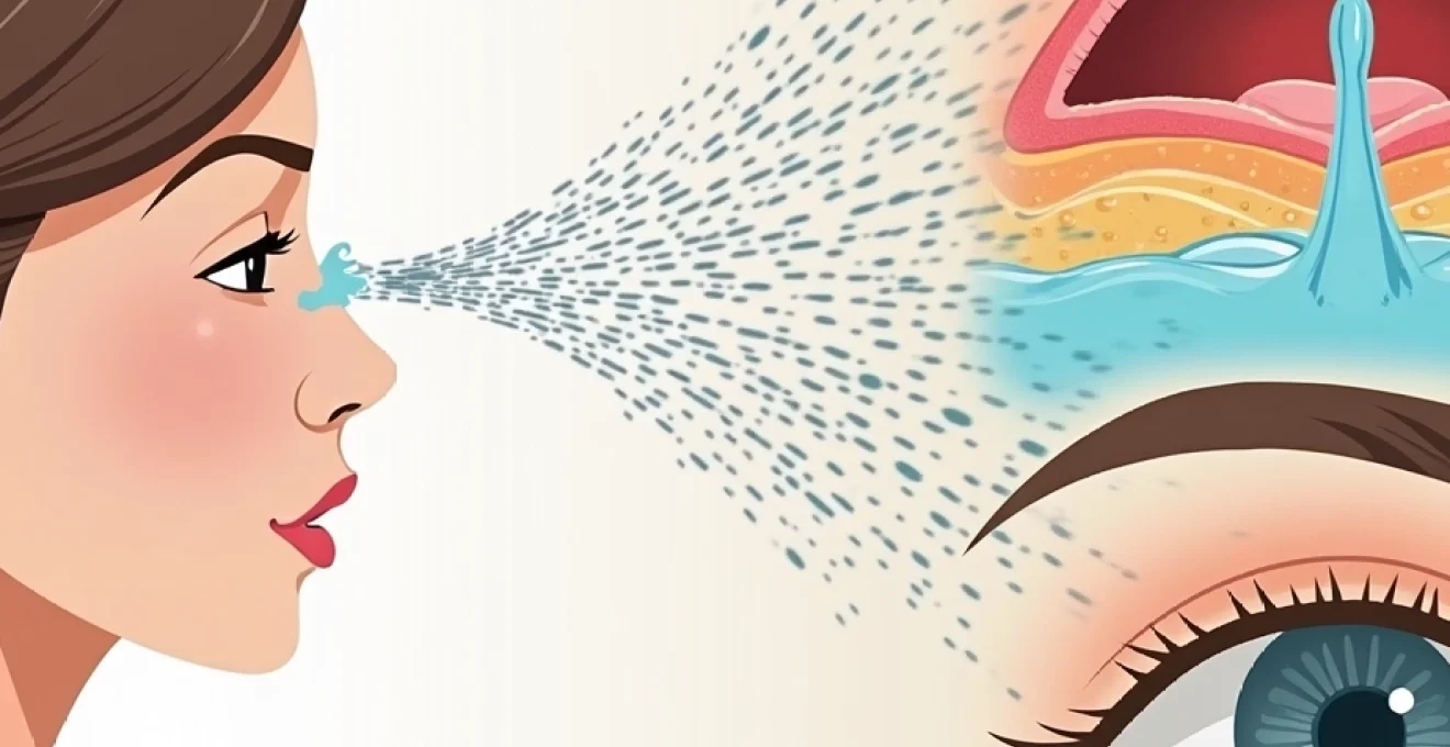
Immediate ocular irrigation protocols for hairspray chemical exposure
The cornerstone of emergency treatment for hairspray eye exposure involves immediate and thorough irrigation of the affected eye. Time becomes critical in these situations, as the longer chemical irritants remain in contact with ocular tissues, the greater the potential for damage. The primary goal of irrigation is to dilute and remove the hairspray components, particularly the alcohol content which typically ranges from 25-50% in most commercial formulations.
Begin irrigation within seconds of exposure, if possible. The affected eye should be held open manually if necessary, as the natural reflex to squeeze eyes shut can trap irritating substances against the corneal surface. Position your head so that the affected eye is lower than the unaffected one, preventing contaminated flush water from flowing into the healthy eye. This positioning becomes particularly important when dealing with bilateral exposures or when helping someone else manage their injury.
Sterile saline solution application techniques for aerosol propellant removal
Sterile saline solution represents the gold standard for emergency ocular irrigation, offering the optimal pH balance and osmolarity for eye tissues. When available, use commercially prepared sterile saline specifically designed for wound irrigation or contact lens care. Pour the solution steadily across the eye surface, directing the flow from the inner corner (nasal side) toward the outer corner (temporal side) to facilitate complete coverage and drainage.
The irrigation should continue for a minimum of 15-20 minutes for most hairspray exposures, though severe cases may require longer flushing periods. Pay particular attention to the lower and upper eyelid margins, as hairspray residue can accumulate in these areas. Propellant chemicals like isobutane can cause additional thermal effects, making thorough removal essential for preventing secondary tissue damage.
Tap water emergency flushing methods when commercial eye wash is unavailable
When sterile saline is not immediately available, clean tap water serves as an acceptable alternative for emergency irrigation. The water should be lukewarm rather than hot or cold, as temperature extremes can cause additional discomfort and potentially worsen tissue irritation. Use a gentle, steady stream rather than high pressure, which could force particles deeper into the eye or cause mechanical trauma to already irritated tissues.
Several practical methods can facilitate effective tap water irrigation. A clean cup filled with water allows for controlled pouring, while shower heads set to gentle pressure provide continuous flow. Kitchen sink sprayers offer good control but should be tested first to ensure appropriate pressure levels. The key principle remains consistent: maintain flow for at least 15-20 minutes while keeping the eye open and allowing thorough drainage.
Morgan lens continuous irrigation systems for severe chemical burns
In healthcare settings, Morgan lenses provide the most effective method for continuous ocular irrigation following severe chemical exposures. These specialised devices consist of a curved plastic shell that fits over the cornea, connected to tubing that delivers a steady stream of irrigating solution. The lens design ensures consistent contact time between the irrigation fluid and all ocular surfaces whilst maintaining patient comfort during extended treatment periods.
Morgan lens systems typically deliver 1-2 litres of normal saline per hour, providing superior irrigation compared to manual methods. The continuous flow helps prevent the reaccumulation of chemical residues and maintains optimal hydration of corneal tissues during the healing process. While primarily used in emergency departments and ophthalmology clinics, understanding their application helps patients recognise when they might encounter this treatment method during professional care.
Contact lens removal procedures during active hairspray contamination
Contact lens wearers face additional complications when hairspray enters their eyes, as the lenses can trap chemical irritants against the corneal surface. Immediate lens removal becomes essential, even though the natural instinct might be to avoid touching irritated eyes. The trapped chemicals can cause concentrated damage to the area directly beneath the lens, potentially leading to more severe corneal injury.
Remove contact lenses immediately after beginning irrigation, not before. The initial flush helps dilute surface contaminants and may make lens removal more comfortable. Use clean hands and standard removal techniques, though some discomfort is expected. If the lens appears stuck or cannot be removed easily, continue irrigation and seek immediate professional help rather than forcing removal, which could cause additional mechanical trauma to already compromised tissues.
Toxicological assessment of common hairspray ingredients affecting ocular tissue
Understanding the specific components in hairspray formulations helps predict the likely severity and type of ocular reaction you might experience. Modern hairsprays contain a complex mixture of polymers, solvents, propellants, and conditioning agents, each with distinct toxicological properties when they contact eye tissues. The concentration and specific combination of these ingredients varies significantly between brands and product types, influencing both immediate symptoms and potential long-term effects.
The most concerning components from an ocular safety perspective include denatured alcohol, which can cause immediate burning and tissue desiccation, and various polymer resins that may cause mechanical irritation or allergic reactions. Propellant gases, while generally less toxic than older chlorofluorocarbon formulations, can still cause thermal effects and pressure-related injuries if the spray nozzle was positioned close to the eye during exposure.
Denatured alcohol concentration effects on corneal epithelium
Denatured alcohol serves as the primary solvent in most hairspray formulations, typically comprising 25-50% of the total product volume. This concentration range places hairspray alcohol content on par with distilled spirits, explaining why accidental ingestion can cause intoxication symptoms. When alcohol contacts the corneal epithelium, it causes immediate cellular dehydration and can disrupt the protective tear film that normally lubricates and protects the eye surface.
The severity of alcohol-induced corneal damage correlates directly with concentration and contact time. Higher alcohol concentrations can cause epithelial cell death, leading to punctate keratitis or larger areas of corneal erosion. These changes typically manifest as severe pain, photophobia, and foreign body sensation. Prompt irrigation remains crucial for limiting alcohol penetration into deeper corneal layers and minimising the extent of epithelial damage.
Polymeric resin particles and conjunctival inflammatory response
Polymeric resins like polyvinylpyrrolidone and carboxymethylcellulose create the holding power in hairsprays, forming a flexible film on hair strands after solvent evaporation. When these polymers contact ocular tissues, they can trigger both mechanical and chemical irritation responses. The particles may become embedded in the conjunctival fornices, creating ongoing irritation even after initial irrigation attempts.
Conjunctival inflammation from polymer exposure typically presents as redness, swelling, and increased tear production. Some individuals may develop delayed hypersensitivity reactions to specific polymer types, resulting in prolonged symptoms that persist beyond the expected recovery period for simple chemical irritation. These reactions may require topical anti-inflammatory treatment and more aggressive mechanical removal of residual particles.
Hydrofluorocarbon propellant thermal burns on periorbital skin
Modern hairspray propellants, primarily hydrofluorocarbons like isobutane, can cause thermal injuries when released at close range to the eye area. The rapid expansion and evaporation of liquefied gas creates a significant cooling effect, potentially causing frostbite-like injuries to periorbital skin and even the corneal surface if exposure is severe enough.
Thermal burns from propellant exposure typically appear as white or grey patches on the skin, accompanied by numbness or severe pain. The eyelids and eyebrows are particularly vulnerable due to their thin skin and proximity to the spray pattern. Cold-induced injuries require different treatment approaches than chemical burns, often necessitating gradual rewarming and careful monitoring for tissue viability. These thermal effects explain why close-range hairspray exposure can cause more severe injuries than might be expected from the chemical components alone.
Fragrance allergen Cross-Reactivity in sensitive individuals
Hairspray fragrances contain multiple allergenic compounds that can trigger immediate or delayed hypersensitivity reactions in susceptible individuals. Common fragrance allergens include limonene, linalool, and various synthetic musks, which may cause contact dermatitis on periorbital skin or allergic conjunctivitis affecting the eye itself. Cross-reactivity between different fragrance families means that individuals with known perfume or cosmetic allergies face increased risk of severe reactions to hairspray exposure.
Allergic reactions typically develop 12-48 hours after initial exposure, distinguishing them from immediate chemical irritation responses. Symptoms may include persistent redness, swelling, and itching that worsens rather than improves with time. Recognition of allergic components becomes important for preventing future exposures and may influence treatment decisions, as allergic reactions often require antihistamine or corticosteroid therapy rather than simple supportive care.
Clinical ophthalmological examination following hairspray exposure
Professional ophthalmological evaluation becomes necessary when symptoms persist beyond initial first aid measures or when certain high-risk features are present. A comprehensive examination typically begins with visual acuity testing to establish baseline function and detect any immediate vision changes. Even minor alterations in visual acuity can indicate corneal epithelial disruption or significant conjunctival swelling that requires medical intervention.
The examination proceeds with external inspection of the eyelids and periorbital area, looking for signs of chemical burns, allergic reactions, or thermal injury from propellant exposure. Fluorescein staining reveals corneal epithelial defects that might not be visible with standard examination techniques. This specialized dye highlights areas where the protective epithelial layer has been damaged, appearing as bright green patches under blue light examination.
Intraocular pressure measurement becomes important in cases of significant exposure, as chemical irritation can sometimes trigger acute angle-closure glaucoma in predisposed individuals. The anterior chamber is examined for signs of inflammation, including cell and flare that indicate breakdown of the blood-aqueous barrier. Conjunctival injection patterns help distinguish between chemical irritation, allergic reactions, and infectious complications that might develop secondarily.
Detailed documentation of findings guides treatment decisions and provides baseline information for monitoring recovery progress. Photography may be used to document the extent of visible damage, particularly useful when multiple follow-up appointments are anticipated. The ophthalmologist will also assess the need for specialist referral, particularly in cases involving significant corneal damage or thermal burns that might require reconstructive interventions.
Professional medical intervention thresholds for hairspray eye injuries
Several specific criteria indicate when hairspray eye exposure requires immediate professional medical attention rather than home management alone. Vision changes of any type, including blurring, double vision, or light sensitivity that doesn’t improve with irrigation, warrant emergency evaluation. These symptoms may indicate corneal damage, intraocular inflammation, or pressure changes that could threaten long-term visual function if left untreated.
Severe pain that persists or worsens after 30 minutes of irrigation suggests significant tissue damage requiring professional intervention. Pain levels that prevent normal eye opening or cause systemic symptoms like nausea indicate more serious injury than typical superficial irritation. Additionally, any visible whitening of the corneal surface suggests epithelial death and potential deeper penetration of chemical agents.
The proximity of the spray nozzle to the eye at the time of exposure significantly influences injury severity, with close-range exposures more likely to cause thermal damage from propellant gases and higher concentrations of chemical irritants.
Patients with pre-existing eye conditions face lower thresholds for seeking professional care. Those with dry eye syndrome, previous corneal surgery, or chronic inflammatory conditions may experience more severe reactions and slower healing times. Contact lens wearers who cannot remove their lenses after irrigation, or who experience persistent foreign body sensation after lens removal, should receive prompt ophthalmological evaluation to rule out lens-related complications or embedded debris.
Signs of allergic reaction development, including progressive swelling, hives around the eye area, or systemic symptoms like difficulty breathing, require immediate medical attention. These reactions can escalate rapidly and may require emergency treatment with antihistamines, corticosteroids, or even epinephrine in severe cases. The combination of chemical injury and allergic reaction creates a complex clinical scenario that demands professional management.
Post-exposure ocular recovery management and complications prevention
Effective recovery management begins immediately after initial irrigation and continues throughout the healing process. The corneal epithelium typically regenerates within 24-48 hours for minor injuries, but this timeline can extend significantly with more severe exposures or in individuals with compromised healing responses. Symptom monitoring becomes crucial during the first 72 hours, as delayed complications can develop even when initial symptoms seem mild.
Environmental modifications support healing and prevent secondary complications. Avoiding windy conditions, dusty environments, and low humidity settings helps maintain corneal hydration and prevents additional irritation. Sunglasses provide protection against photophobia while supporting the natural healing process by reducing light-induced discomfort that can interfere with blinking patterns necessary for tear film distribution.
Activity restrictions may be necessary during the acute recovery phase. Swimming, particularly in chlorinated pools, should be avoided until epithelial healing is complete. Contact lens wear requires careful consideration and often temporary discontinuation to prevent mechanical irritation of healing tissues. Makeup application around the eye area should be postponed until all symptoms resolve to avoid introducing additional chemical irritants or bacterial contamination.
Recovery timelines vary significantly based on individual factors including age, overall health status, and pre-existing ocular conditions, with complete healing typically occurring within one to two weeks for most minor exposures.
Antibiotic prophylaxis protocols for secondary bacterial infections
Secondary bacterial infections represent a significant risk following chemical eye injuries, as damaged epithelial barriers provide entry points for pathogenic organisms. Prophylactic antibiotic use remains controversial, with most ophthalmologists reserving topical antibiotics for cases with significant epithelial disruption or high-risk patient populations. When prescribed, broad-spectrum topical antibiotics like polymyxin B-trimethoprim or fluoroquinolones provide coverage against common ocular pathogens.
Signs of developing infection include increasing pain after initial improvement, purulent discharge, and progressive redness despite appropriate supportive care. These symptoms typically develop 2-5 days after initial injury and require prompt antibiotic treatment to prevent serious complications like corneal perforation or endophthalmitis. Patient education about infection warning signs becomes crucial for early detection and intervention.
Artificial tear supplementation schedules for dry eye syndrome
Artificial tear supplementation plays a vital role in supporting corneal healing and preventing secondary dry eye syndrome following hairspray exposure. The chemical components in hairspray can disrupt normal tear film composition and reduce natural tear production, creating ongoing discomfort even after initial tissue healing is complete. Preservative-free formulations are preferred, as preserved drops can cause additional irritation in already compromised eyes.
Application schedules typically begin with frequent dosing every 1-2 hours during waking hours for the first 24-48 hours, then gradually reducing frequency as symptoms improve. Gel-based artificial tears provide longer-lasting relief but may cause temporary visual blurring, making them more suitable for bedtime use. Some patients benefit from combination therapy using different viscosity products at different times of day to optimise both comfort and visual function.
Corneal abrasion healing monitoring using fluorescein staining
Fluorescein staining provides objective assessment of corneal epithelial healing progress and helps guide treatment modifications throughout recovery. Initial staining patterns document the extent and location of epithelial damage, while serial examinations track healing rates and identify areas of delayed recovery that might require additional intervention. Complete epithelial healing typically occurs within 24-72
hours for minor injuries, though more extensive damage may require up to a week for complete resolution.
Regular follow-up examinations allow for treatment adjustments based on healing progress. Patients showing delayed healing or persistent epithelial defects may benefit from additional interventions such as bandage contact lenses, which provide mechanical protection and maintain corneal hydration. Punctal occlusion techniques can help conserve natural tears in cases where dry eye symptoms persist beyond expected recovery timeframes.
The characteristic bright green fluorescence indicates areas where epithelial cells have been lost or damaged, creating defects in the protective barrier. As healing progresses, these areas gradually diminish in size and intensity until complete epithelial coverage is restored. Documentation of healing patterns helps identify factors that might be impeding recovery, such as incomplete removal of chemical residues, developing infections, or underlying conditions affecting epithelial regeneration rates.
Advanced imaging techniques like anterior segment optical coherence tomography provide detailed cross-sectional views of corneal structure during healing, particularly useful in cases involving deeper tissue damage. These technologies allow for precise measurement of epithelial thickness and identification of subtle structural changes that might not be apparent with conventional examination methods. Understanding the relationship between clinical symptoms and objective healing markers helps both patients and healthcare providers maintain realistic expectations about recovery timelines.
Modern ophthalmological monitoring techniques can detect healing progress at the cellular level, allowing for precise treatment modifications that optimise recovery outcomes while minimising the risk of long-term complications.
Patient education about normal versus abnormal healing patterns empowers individuals to recognise when additional medical attention becomes necessary. Most patients experience gradual improvement in pain levels and light sensitivity over the first 48-72 hours, with complete symptom resolution typically occurring within one to two weeks. Deviations from this expected pattern, particularly worsening symptoms after initial improvement, warrant prompt ophthalmological evaluation to rule out complications such as infection, persistent chemical residue, or allergic reactions requiring modified treatment approaches.