Four months following anterior cervical discectomy and fusion (ACDF) surgery represents a critical milestone in your recovery journey. At this stage, most patients experience significant improvements in their neurological symptoms while continuing to adapt to the structural changes within their cervical spine. The fusion process has typically advanced considerably, with solid bone formation beginning to establish permanent stability between the treated vertebral segments.
This period marks a transition from the acute healing phase to long-term recovery, where you can begin to assess the true effectiveness of your surgical intervention. Many patients find themselves cautiously optimistic as chronic pain diminishes and mobility gradually returns. However, understanding what constitutes normal progress versus concerning developments remains crucial for optimal outcomes and patient confidence during this extended recovery period.
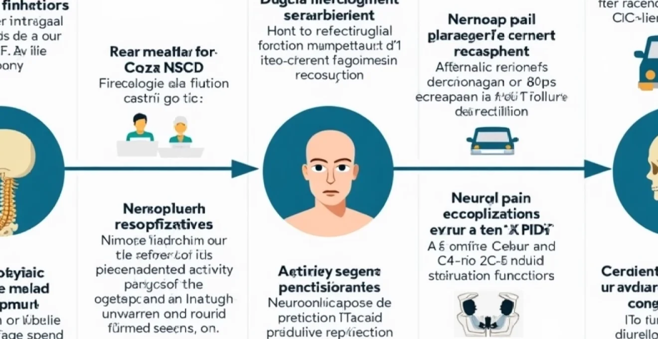
Physical recovery milestones at Four-Month Post-ACDF timeline
The four-month mark represents a pivotal assessment period where surgeons can evaluate the success of cervical fusion and overall healing progress. Most patients demonstrate substantial improvements in their pre-operative symptoms, including reduced radicular pain, improved strength, and enhanced functional capacity. The inflammatory response that characterised the initial healing phase has typically subsided, allowing for more accurate evaluation of surgical outcomes.
Physical recovery at this stage involves multiple interconnected systems working together to restore optimal cervical spine function. Your body has spent considerable energy remodelling bone tissue, adapting muscular patterns, and re-establishing neural pathways that may have been compromised by pre-operative compression. The integration of these healing processes determines your overall functional improvement and long-term prognosis.
Cervical spine fusion assessment through CT and X-Ray imaging
Advanced imaging studies at four months post-surgery provide crucial insights into fusion progression and structural integrity. Computed tomography scans offer superior bone detail, allowing surgeons to evaluate trabecular bridging and cortical continuity across the fusion construct. Plain radiographs, while less detailed, remain valuable for assessing overall alignment and hardware positioning during dynamic movements.
Fusion assessment criteria include evidence of solid bone formation between vertebral bodies, absence of motion on flexion-extension radiographs, and appropriate integration of bone graft materials. Successful fusion typically demonstrates continuous trabecular patterns extending from one vertebra to another, indicating mechanical stability and biological incorporation. Some patients may show incomplete fusion at four months, which doesn’t necessarily indicate surgical failure, as fusion can continue progressing for up to twelve months post-operatively.
Range of motion recovery in C5-C6 and C6-C7 vertebral segments
Range of motion recovery varies significantly depending on the specific levels treated and individual patient factors. Single-level fusions, particularly at C5-C6 or C6-C7, typically preserve substantial overall cervical mobility due to compensation from adjacent segments. Multi-level procedures naturally result in more significant motion restrictions, though functional limitations are often minimal for daily activities.
Cervical rotation, lateral flexion, and sagittal plane movements should demonstrate measurable improvement compared to pre-operative restrictions caused by pain and muscle spasm. The cervical spine’s remarkable adaptability allows adjacent segments to gradually accommodate for fused levels, though this adaptation process may contribute to accelerated degeneration over time. Most patients report functional satisfaction with their range of motion by four months, particularly when pre-operative symptoms have resolved.
Neurological function restoration following anterior decompression
Neurological recovery represents one of the most significant benefits of ACDF surgery, with substantial improvements typically evident by four months post-operatively. Radicular symptoms, including arm pain, numbness, and tingling, show marked improvement in approximately 85-95% of patients. Myelopathic symptoms, such as hand clumsiness, gait instability, and weakness, may continue improving for six to twelve months following decompression.
Nerve root recovery follows predictable patterns, with sensory symptoms often improving before motor function. The extent of neurological recovery correlates closely with the duration and severity of pre-operative compression. Patients with acute onset symptoms generally experience more complete recovery compared to those with chronic, progressive neurological deficits. Electromyography studies may demonstrate ongoing nerve regeneration even when clinical symptoms have plateaued.
Muscular strength recovery in cervical paraspinal and trapezius groups
Muscular strength recovery encompasses both the restoration of previously weakened muscles and adaptation to altered biomechanics following fusion. Deep cervical flexors, which often become inhibited due to pre-operative pain, require specific attention during rehabilitation. Trapezius and levator scapulae muscles may demonstrate improved function as neural compression resolves and normal activation patterns return.
Physical therapy protocols at four months typically emphasise progressive strengthening exercises targeting both local stabilisers and global mobilisers of the cervical spine. Coordinated muscle activation patterns are essential for maintaining cervical stability and preventing compensatory movement strategies that could predispose to adjacent segment problems. Most patients demonstrate measurable strength improvements in previously affected muscle groups by this recovery milestone.
Pain management and medication tapering protocols
Pain management at four months post-ACDF surgery focuses on transitioning from acute pain control to long-term comfort maintenance. Most patients experience substantial reduction in their pre-operative pain levels, though some residual discomfort may persist as tissues continue healing and adapting. The inflammatory phase has typically resolved, allowing for more targeted approaches to managing specific pain generators.
Medication requirements generally decrease significantly by this timepoint, with many patients reducing or eliminating prescription pain medications entirely. However, individualised approaches remain essential as some patients may require ongoing pharmaceutical support for optimal comfort and function. The goal shifts from controlling acute post-surgical pain to managing any persistent symptoms while minimising medication dependence and side effects.
Transitioning from Opioid-Based to NSAIDs pain control regimens
Opioid tapering should be well advanced by four months post-surgery, with most patients successfully transitioning to non-opioid alternatives or achieving complete discontinuation. Non-steroidal anti-inflammatory drugs (NSAIDs) become more appropriate for long-term use once the critical bone healing phase has passed, though some concerns about fusion inhibition may persist depending on individual risk factors.
Gradual opioid reduction protocols help prevent withdrawal symptoms while maintaining adequate pain control during the transition period. Alternative medications such as acetaminophen, topical analgesics, and muscle relaxants may provide sufficient symptom management for most patients. Those requiring continued opioid therapy should undergo comprehensive evaluation to identify persistent pain generators and optimise treatment approaches.
Neuropathic pain resolution through gabapentin and pregabalin adjustment
Anticonvulsant medications used for neuropathic pain management often require dose adjustments as nerve compression resolves and symptoms improve. Gabapentin and pregabalin doses may be gradually reduced or discontinued entirely as radicular symptoms resolve. Some patients continue benefiting from low-dose neuropathic agents for residual symptoms or as preventive therapy.
Neuropathic pain resolution typically follows nerve healing patterns, with improvements continuing for several months following successful decompression. Medication titration should be guided by symptom response rather than arbitrary timelines, as individual recovery rates vary considerably. Patients experiencing persistent neuropathic symptoms may require alternative medications or additional diagnostic evaluation to identify ongoing compression or other pain generators.
Muscle relaxant discontinuation following baclofen and cyclobenzaprine usage
Muscle relaxants prescribed for post-operative muscle spasm and tension can typically be discontinued by four months as normal muscle function returns. Baclofen, cyclobenzaprine, and similar medications should be gradually tapered to prevent rebound muscle tension and withdrawal effects. Most patients find that improved biomechanics and resolved nerve irritation eliminate the need for ongoing muscle relaxant therapy.
Progressive exercise and physical therapy provide more sustainable approaches to maintaining muscle relaxation and preventing tension-related symptoms. Some patients may benefit from occasional use of muscle relaxants during periods of increased activity or stress, but daily use should generally be unnecessary by this recovery stage.
Alternative pain management through cervical epidural steroid injections
Cervical epidural steroid injections may be considered for patients experiencing persistent radicular symptoms or inflammation around the surgical site. These procedures can provide targeted anti-inflammatory effects while avoiding systemic medication side effects. Timing of injections relative to surgery requires careful consideration to avoid interference with fusion processes.
Injection therapy success rates vary depending on the underlying pathology and technical factors related to needle placement and medication selection. Fluoroscopic or CT-guided approaches ensure accurate medication delivery to affected nerve roots or epidural spaces. Multiple injection sessions may be required for optimal benefit, though the need for repeated procedures should prompt evaluation for other pain sources or surgical complications.
Activity restrictions and gradual return to normal function
Activity progression at four months post-ACDF surgery allows for substantial expansion of permitted movements and functional tasks. Most lifting restrictions can be liberalised, though heavy manual labour may still require modifications or alternative techniques. Sports participation becomes possible for many activities, though contact sports and high-impact exercises typically remain contraindicated until fusion is completely solid.
The gradual return to normal function requires balancing the desire for activity with the need to protect the healing fusion construct. Progressive loading principles guide activity advancement, allowing tissues to adapt gradually to increasing demands. Most patients find that their functional capacity exceeds their pre-operative abilities as pain resolves and neurological function improves, though some activities may require permanent modifications due to altered cervical mechanics.
Work-related activities typically resume with minimal restrictions by four months, though job-specific considerations may require ongoing accommodations. Desk workers generally return to full duties, while manual labourers may need ergonomic assessments and task modifications to prevent excessive stress on the cervical spine. Driving restrictions are usually lifted, though some patients experience continued discomfort with prolonged sitting or repetitive neck movements.
Exercise protocols emphasise cardiovascular fitness, core strengthening, and gradual return to recreational activities. Swimming provides excellent low-impact exercise, though diving and high-impact water activities should be avoided. Walking, cycling, and elliptical training offer safe cardiovascular options while minimising cervical spine stress. Individual tolerance and surgeon approval should guide specific exercise selection and progression.
Potential complications and warning signs at Four-Month mark
While most patients experience successful outcomes by four months post-ACDF surgery, potential complications require ongoing vigilance and prompt recognition. Early identification of problems allows for timely intervention and improved long-term outcomes. Understanding warning signs empowers patients to seek appropriate medical attention when concerning symptoms develop.
Complications at this stage may represent delayed manifestations of surgical issues or new problems related to healing processes and biomechanical changes. Distinguishing normal recovery variations from pathological developments requires clinical expertise and appropriate diagnostic studies. Regular follow-up appointments and patient education remain crucial components of comprehensive post-operative care.
Pseudoarthrosis development and Non-Union risk assessment
Pseudoarthrosis, or failure of bone fusion, represents one of the most significant potential complications following ACDF surgery. Risk factors include smoking, diabetes, osteoporosis, multi-level procedures, and certain medications that impair bone healing. Clinical symptoms may include recurrent neck pain, radicular symptoms, or mechanical symptoms suggesting motion at the fusion site.
Diagnostic evaluation for pseudoarthrosis typically includes flexion-extension radiographs to assess for abnormal motion, CT scans to evaluate bone formation, and potentially bone scan studies to assess biological activity. Treatment options for confirmed pseudoarthrosis may include revision surgery with bone grafting, electrical bone stimulation, or alternative fusion techniques depending on specific circumstances and patient factors.
Adjacent segment disease in C4-C5 and C7-T1 levels
Adjacent segment disease represents accelerated degeneration of spinal levels immediately above or below the fused segments. This phenomenon results from altered biomechanics that increase stress on adjacent motion segments. Symptoms typically include neck pain, radicular symptoms, or myelopathic signs similar to the original pathology but originating from different anatomical levels.
The incidence of adjacent segment disease increases over time, with some studies reporting rates of 10-25% within five to ten years following cervical fusion. Prevention strategies include maintaining proper cervical alignment and avoiding unnecessary multi-level fusions when possible. Treatment approaches mirror those for primary cervical pathology, though revision surgery carries increased complexity and potential complications.
Hardware-related complications including cage migration and screw loosening
Hardware complications may manifest as cage migration, screw loosening, or plate-related problems affecting adjacent structures. Symptoms can include recurrent pain, neurological deficits, or mechanical symptoms suggesting hardware displacement. Regular radiographic monitoring helps identify these complications before they become symptomatic or cause permanent damage.
Risk factors for hardware complications include inadequate bone quality, technical factors related to implant placement, and excessive mechanical stress before fusion consolidation. Modern implant designs and surgical techniques have reduced complication rates, though vigilance remains necessary throughout the healing period. Treatment typically requires revision surgery to address displaced or failed hardware components.
Long-term prognosis and quality of life outcomes
Long-term prognosis following ACDF surgery is generally excellent, with success rates consistently reported between 85-95% for appropriately selected patients. Quality of life improvements typically exceed pre-operative expectations as pain resolves and neurological function returns. Most patients report satisfaction with their surgical outcomes and would choose surgery again if faced with similar circumstances.
Functional outcomes demonstrate sustained improvements in activities of daily living, work capacity, and recreational pursuits. The resolution of chronic pain and neurological symptoms often has profound psychological benefits, including improved mood, sleep quality, and overall life satisfaction. Many patients describe feeling “like themselves again” as debilitating symptoms that may have persisted for months or years finally resolve.
Durability of surgical results extends well beyond the immediate post-operative period, with studies demonstrating maintained benefits at five, ten, and fifteen-year follow-up intervals. While some patients may develop adjacent segment disease or other age-related spinal changes, these complications typically occur years after the initial surgery and may not require additional intervention. The vast majority of patients maintain their improved quality of life indefinitely following successful ACDF procedures.
Follow-up care requirements and imaging schedule protocols
Ongoing follow-up care ensures optimal outcomes and early detection of potential complications throughout the recovery period and beyond. Regular appointments allow for assessment of fusion progression, symptom resolution, and functional improvement while providing opportunities to address patient concerns and adjust treatment plans as needed. Structured follow-up protocols have been shown to improve patient satisfaction and clinical outcomes.
Imaging schedules typically include plain radiographs at regular intervals to assess fusion progression and hardware positioning, with advanced imaging reserved for specific clinical indications. CT scans may be obtained at six to twelve months to confirm solid fusion, while MRI studies are generally reserved for patients with persistent or recurrent symptoms suggesting complications or adjacent segment pathology. The timing and frequency of imaging studies should be individualised based on clinical progress and risk factors.
Long-term surveillance focuses on maintaining cervical spine health and preventing future problems through lifestyle modifications, ergonomic awareness, and appropriate exercise programs. Annual or biennial follow-up appointments may be sufficient for asymptomatic patients with confirmed fusion, though more frequent monitoring may be necessary for those with risk factors for complications or adjacent segment disease. Patient education regarding warning signs and when to seek medical attention remains a crucial component of comprehensive long-term care strategies.